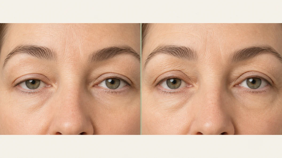
“My eyes look tired and hooded” can describe at least two genuinely different anatomical problems, and mistaking one for the other is an avoidable source of disappointment in eyelid surgery. Getting the diagnosis right before a surgical plan is made matters more than comparing generic before-and-after photographs.
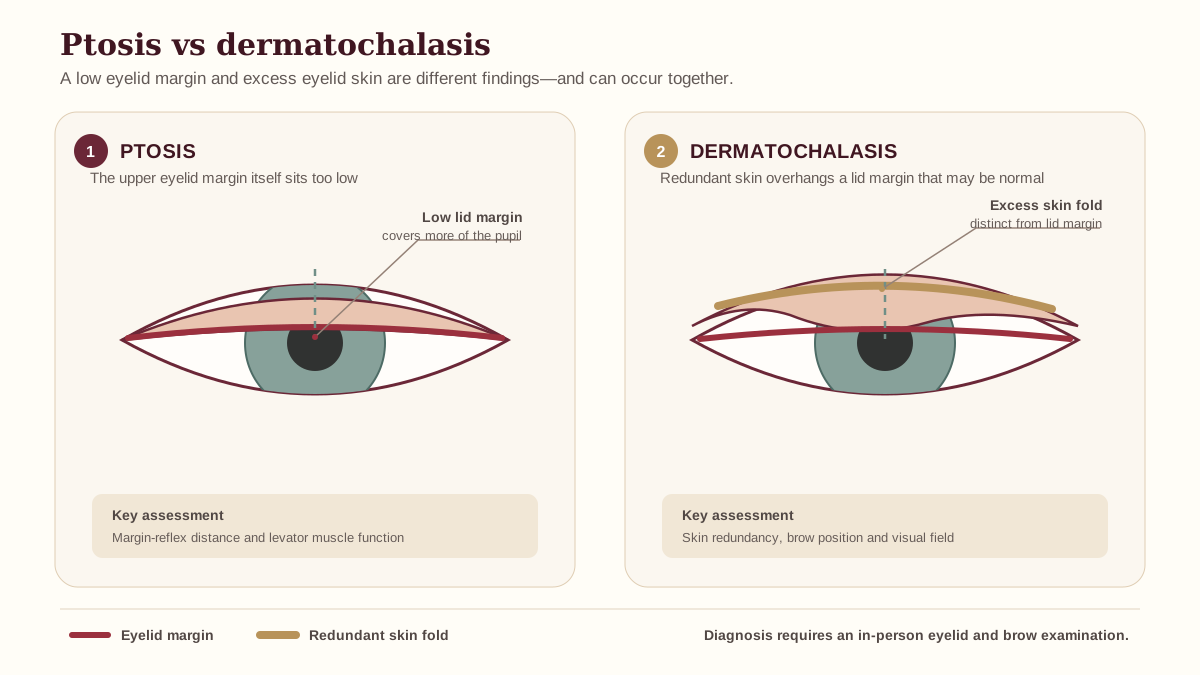
Dermatochalasis — Excess Upper Eyelid Skin
Dermatochalasis refers to loose, redundant skin on the upper eyelid — typically developing gradually with age as skin loses collagen and elasticity. The eyelid margin itself (where the lash line sits) remains at a normal anatomical position; it's the skin folding over it that creates the hooded, heavy appearance. The problem is purely one of skin laxity, and it usually progresses with each decade, often becoming more symptomatic from the 40s or 50s onward.
This is the condition that standard upper blepharoplasty is designed to treat — the procedure removes the excess skin and, where present, the herniated fat pad contributing to puffiness. It's a well-understood operation with predictable outcomes when the diagnosis is accurate. Patient satisfaction after upper blepharoplasty for documented dermatochalasis is consistently high, including measurable functional improvement in superior visual field.1
Ptosis — a Drooping Eyelid Margin
Ptosis is a fundamentally different problem: the eyelid margin itself sits too low, typically because the levator muscle — the primary mechanism responsible for lifting the eyelid — and its aponeurosis (the tendon connecting it to the tarsal plate) have weakened, stretched, or partially detached. This most commonly occurs with age (involutional ptosis) as the levator aponeurosis gradually disinserts. Other causes include prior intraocular surgery, extended contact lens use, and — less commonly — neuromuscular conditions or a congenital origin.
Because the problem is muscular and tendinous, removing skin does nothing to fix it. Ptosis requires a surgical repair that specifically tightens or reattaches the levator mechanism — a different operation from standard blepharoplasty, performed by surgeons with oculoplastic or specific eyelid training. In some cases, the Müller's muscle (a secondary eyelid elevator) is addressed instead of or alongside the levator, depending on the degree and type of ptosis identified on examination.
Why Misdiagnosis Leads to Disappointing Results
Because both conditions can produce a similarly hooded, heavy-looking eye from the outside, it's genuinely possible for a patient — or, in less careful hands, a surgeon — to assume the problem is simply excess skin when true ptosis is actually present or contributing. If only skin is removed and underlying ptosis is left uncorrected, the eye can still look droopy after healing — sometimes to the patient's genuine confusion, since they've already had surgery. This is one of the most common, and most avoidable, sources of poor outcomes in elective eyelid surgery.
| Feature | Dermatochalasis | Ptosis |
|---|---|---|
| Problem location | Excess overlying skin | Low eyelid margin (levator mechanism) |
| Eyelid margin position (MRD) | Normal | Reduced (low relative to pupil) |
| Treatment | Upper blepharoplasty (skin removal) | Levator repair / ptosis procedure |
| Can they coexist? | Yes — and they frequently do, especially in older adults | |
| Functional visual field impact | Yes, from overhanging skin | Yes, from low lid position |
| Result if wrong op performed | — | Persistent droop despite blepharoplasty |
The implication for patients is clear: a proper eyelid examination before any surgical plan is finalized is not optional. A consultation that focuses on your desired outcome without specifically measuring eyelid margin position is missing the diagnostic step that determines which surgery — or which combination — is actually indicated.
How It's Diagnosed
A proper eyelid examination goes beyond visual impression. The key objective measurement is the margin reflex distance (MRD) — specifically, how far the upper eyelid margin sits above the center of the pupil (the corneal light reflex) when the patient looks straight ahead in primary gaze. A normal MRD-1 is approximately 4 to 5 mm; values of 2 mm or less indicate significant ptosis; values between 2 and 4 mm may reflect mild to moderate ptosis that still warrants treatment.
The examination also assesses levator function — how far the eyelid travels from downgaze to upgaze, which directly reflects the integrity of the levator mechanism. Normal levator excursion is typically 15 mm or more; values below 10 mm suggest poorer levator function and may change which repair approach is most appropriate. In cases where the diagnosis is unclear, the phenylephrine test (instilling a topical sympathomimetic to stimulate Müller's muscle) can help predict whether a Müller's-only procedure will be sufficient.
Many patients, particularly older adults, have a genuine mix of both dermatochalasis and involutional ptosis simultaneously — which is why an experienced examiner will assess for both rather than stopping at the more obvious finding. Combined surgery addressing skin removal and levator repair in the same operative session is a standard approach when both conditions are present.
Brow Position as a Third Factor
A third variable that complicates the picture is brow position. A heavy or low-set brow can rest directly on the upper eyelid, creating a hooded appearance that visually mimics dermatochalasis or ptosis — but is actually being driven, or substantially worsened, by the brow itself. Surgeons use the term brow ptosis for this condition: the brow resting at or below the supraorbital rim, which is below its anatomically normal position in the relaxed face.
When brow ptosis is the primary driver, eyelid surgery alone may under-deliver — because the excess skin the blepharoplasty removes will be partly replaced by the continuing descent of the heavy brow onto the lid. This is a common reason for patients to be disappointed after technically well-performed blepharoplasty. A brow lift may need to be part of the discussion before any eyelid surgery is planned. See our brow lift results guide for more on this overlap and when the combination makes sense.
Insurance & Visual Field Testing
Whether the underlying issue is dermatochalasis, ptosis, or both, insurance coverage is possible — though not guaranteed — when formal visual field testing documents that the condition is meaningfully obstructing your superior vision. The functional pathway for both conditions follows similar documentation logic: a published Goldmann perimetry study of upper blepharoplasty patients found an average 4.99-fold improvement in superior visual field after surgery, providing the kind of objective measurement that supports functional coverage claims.2
In practice, insurers typically require: formal visual field testing (Goldmann or automated perimetry) both without and with the lids taped up to simulate correction, demonstrating a meaningful difference; examination notes documenting the degree of lid/skin overhang; and often a trial period of conservative management. The criteria are similar whether the claim is for dermatochalasis or ptosis, though insurers may handle the two CPT codes differently. See our insurance coverage guide for the general documentation requirements that apply to functional eyelid surgery.
Frequently Asked Questions
Do I have ptosis or just extra skin?
Only an eye-focused exam can tell the two apart reliably, since both can make the eyes look tired or hooded from the outside. A clinician measures the position of your upper eyelid margin relative to the pupil (margin reflex distance) to determine whether the lid itself is drooping (ptosis) or whether excess skin is simply overhanging a normally positioned lid (dermatochalasis).
Will blepharoplasty fix a droopy eyelid?
Standard upper blepharoplasty removes excess skin, so it treats dermatochalasis effectively, but it does not correct true ptosis, which involves the muscle and tendon that lift the eyelid itself. If ptosis is present and only skin is removed, the eye may still look droopy after surgery — a separate ptosis repair addresses the lifting mechanism directly.
Is ptosis surgery covered by insurance?
It can be, if visual field testing documents that the drooping eyelid is significantly obstructing your vision, similar to the functional criteria used for blepharoplasty coverage. Coverage always depends on your specific plan and documented exam findings — see our insurance coverage guide for the general criteria insurers apply.
Can I have both ptosis and dermatochalasis at the same time?
Yes, and it's common, especially in older adults — a thorough exam should assess for both conditions rather than assuming only one is present. Combined surgery addressing skin removal and levator repair simultaneously is a standard approach in this scenario.
Does brow position affect this diagnosis?
Yes — a low or heavy brow can create a hooded look that mimics or worsens both conditions, and eyelid surgery alone may not fully resolve the appearance if the brow is the true driver. A brow lift may need to be discussed alongside eyelid surgery in that case.
Who should diagnose ptosis vs dermatochalasis?
An oculoplastic surgeon, ophthalmologist, or plastic surgeon with specific eyelid experience is best positioned to make this distinction accurately using a proper eyelid exam and measurements. This diagnostic step should happen before any surgical plan is agreed upon.